When many people hear the terms assassination and sabotage the first thing that comes to mind is almost certainly a James Bond movie or some other stereotypical spy flick. In this movie, the dashing charismatic lead infiltrates an enemy stronghold to hinder the plans of some nefarious villain who plans to dispose of the hero in unforetold ways. Well, it has often been said that the truth is stranger than fiction and in the case of Leishmania
donvani, it is certainly the case.
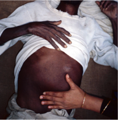 |
Figure 1. Severe
splenomegaly or
enlargement of the spleen due
to visceral leishmaniasis.
|
Every year approximately 500,000 new cases of VL are recorded and approximately 50,000-70,000 deaths occur due to this disease. It has been reported that the only parasitic disease with a higher death toll is malaria. Most of these cases (>90%) have been recorded in Bangladesh, India, Nepal, Sudan, Ethiopia, and Brazil (1, 2). It is disheartening to relay that areas that are endemic to VL are often rural and associated with poverty. Due to this disease families often spend large amounts of money on treatment and often lose income due to the infected individual’s inability to work (1, 2) (Fig. 2).
 |
| Figure 2. Reported new cases of VL according to the WHO as of 2013. A majority of cases occur in Bangladesh, India, Nepal, Sudan, Ethiopia, and Brazil. |
At this point, it is pretty obvious
that Leishmania donovani isn’t the
“good guy” in this scenario. In fact, this parasite is more like an undercover
agent pulling strings from behind the scenes to benefit itself at the expense
of its’ victims. The question is: how did Leishmania
donovani implant itself into humans in order to benefit itself and cause
disease?
Leishmania species are typically transferred between hosts in the gut of a female phlebotomine sand fly. Within
this sand fly, Leishmania are typically in a promastigote form. The promastigote form is characterized by
having long cells that are able to move using a flagellum within the fly (1–3). Within the sand fly Leishmania species replicate and prepare
for the invasion of a new host (2). When the female sand fly
finds an unfortunate individual deemed worthy of her next meal the
promastigotes are transferred into a new human host. Once inside a host, the
promastigotes migrate to the liver. The presence of the parasite activates an
immune response in the human host. One of the first human immune cells that
reacts to this new infection is the macrophage. The macrophage identifies the
promastigote as an invader and tries to surround and destroy it using a process
called phagocytosis. During this process, promastigotes use a variety of
strategies to avoid being killed by the macrophages. Some of these strategies
include preventing the release of deadly antimicrobial agents and the
inhibition of other macrophage functions that are required for an immune
response to the parasite (3). These activities allow
for the survival of the parasite within the macrophage. Differentiation of the
promastigote form into the amastigote form occurs next. The amastigote form is
characterized by the loss of the flagella and round cells (1–3). The amastigotes then
replicate within the disabled macrophage (1). The multiplication and
spread of amastigotes throughout the body is what leads to typical LV symptoms
and systemic infection. After spreading throughout the body another female
phlebotomine sand fly can then become infected and have the amastigotes
differentiate into the promastigote form to repeat the cycle (Fig. 3).
 |
|
Figure 3. The
life cycle of Leishmania species.
Transmission
occurs from female sandflies while in the promastigote form.
Change into the amastigote form occurs in a human host.
Multiplication of
amastigotes leads to characteristic VL symptoms.
|
Dicer
is a protein that normally plays a role in the creation of micro RNAs. A micro
RNA (miR) is an RNA that is created during the transcription of a DNA sequence
to an RNA sequence. Unlike many RNAs transcribed, miRNAs do not code for a
protein sequence and are often referred to as noncoding RNAs. Although these
noncoding RNAs do not play a role in the formation of protein they have been observed
to be involved in post-transcriptional regulation. This means that they play a
role in the regulation of gene expression and help to control the amounts of
proteins that are made from a specific gene. The use of miRNAs in
post-transcriptional regulation starts with the creation of a DNA sequence in
the nucleus that codes for a noncoding miRNA. These newly formed miRNAs are
called pri-miRNAs and form the hairpin loop structures shown in Figure 4 (4). The
newly formed pri-miRNAs interact with a protein complex made up of two proteins
called Drosha and Pasha (aka DGCR8). The protein complex containing Drosha and
Pasha is used to cleave loose hanging pri-miRNA ends and process them so they
are better suited for their jobs. After the interaction with the Drosha and
Pasha protein complex, the miRNAs are referred to as pre-miRNAs. The newly
formed pre-miRNAs still have a hairpin loop structure and are then transported
out of the nucleus where they encounter the Dicer protein. Dicer’s role is to
cleave the loop portion of the hairpin structure and to further fine tune the
pre-miRNAs for their regulatory jobs. After the interaction with Dicer, the
pre-miRNAs are now considered full-fledged miRNAs. These miRNAs are loaded onto
RISC protein complexes. These RISC complexes escort the miRNAs around various
areas in the cell to perform regulation activities. Now that we understand how
Dicer normally functions to help in the creation of miRNAs the assassination of
Dicer and sabotage of cholesterol metabolism can be examined.
 |
|
Figure 4. Mechanism
of Dicer cleavage as described by Descoteaux and colleagues (4). The GP-63
protease cleaves Dicer to prevent the maturation of miR-122 pre-RNA. This
causes a decrease in the levels of cholesterol within the human host. This
allows for Leishmania to replicate
and spread through the body easier.
|
Normally, the regulation of cholesterol metabolism in the
liver is partially controlled by the post-transcriptional control using a miRNA
called miR-122 (5). During infection with Leishmania donovani, a protein called a GP-63 protease has been
shown to be used to cleave Dicer (6). Since Dicer is no longer functional
miR-122 copies can no longer mature or function to regulate cholesterol
metabolism (6). The loss of functional copies of
miR-122 leads to lower levels of cholesterol in the serum of human host. This
has been shown to be beneficial for the invasion of Leishmania donovani and allows for further infection of liver cells
in a human host (7). The next question to ask is how the
GP-63 protease moves from a Leishmania
donovani parasite within a macrophage to a human liver cell to cleave
Dicer. Unfortunately, the process by which this happens is unknown and the
undercover agent still remains behind the scenes of the crime pulling the strings
for its’ own benefit.
Before you leave thinking that all hope is lost
and that the undercover agent will forever remain in the shadows of the
macrophage pulling the strings to our liver cells for eternity let us have a
debrief. The undercover operative Leishmania
donovani is a protozoan parasite. It is also the main causative factor of
the disease visceral leishmaniasis which is primarily observed in Bangladesh, India, Nepal,
Sudan, Ethiopia, and Brazil.
The only reason this disease is able to manifest is due to the stealth and
techniques employed by Leishmania
donovani to evade the immune system and facilitate infection. The most
notable of these techniques is the assassination of Dicer using a GP-63 protease.
This assassination ultimately leads to the sabotage of cholesterol metabolism
within the unsuspecting victim and allows for Leishmania donovani to replicate, spread, and cause disease easier.
Although the process by which this undercover agent performs this assassination
and sabotage is still shrouded in mystery there is no need to fear! There is
still hope that the scientific community will finally discover how to purge
this menace from the depths of unsuspecting macrophages and free us from this disease.
So carry on fellow microbiology enthusiast and fear not the undercover agent
that invades, assassinates, and sabotages for the future only holds more
scientific discovery and with it the information required to expunge the
undercover agent named Leishmania
donovani.
Citations
1. Chappuis F, Sundar S, Hailu A, Ghalib H,
Rijal S, Peeling RW, Alvar J, Boelaert M. 2007. Visceral leishmaniasis: what
are the needs for diagnosis, treatment and control? Nat Rev Microbiol 5:S7–S16.
2. Murray HW, Berman JD, Davies CR, Saravia
NG. 2005. Advances in leishmaniasis. Lancet 366:1561–1577.
3. Olivier M, Gregory DJ, Forget G. 2005.
Subversion mechanisms by which Leishmania parasites can escape the host immune
response: A signaling point of view. Clin Microbiol Rev. American Society for
Microbiology.
4. Descoteaux A, Moradin N, Arango Duque G.
2013. Leishmania dices away cholesterol for survival. Cell Host Microbe
13:245–247.
5. Wen J, Friedman JR. 2012. miR-122
regulates hepatic lipid metabolism and tumor suppression. J Clin Invest
31:2773–2776.
6. Ghosh J, Bose M, Roy S, Bhattacharyya SN.
2013. Leishmania donovani targets dicer1 to downregulate miR-122, lower serum
cholesterol, and facilitate murine liver infection. Cell Host Microbe
13:277–288.
7. Ghosh J, Das S, Guha R, Ghosh D, Naskar
K, Das A, Roy S. 2012. Hyperlipidemia offers protection against Leishmania
donovani infection: role of membrane cholesterol. J Lipid Res 53:2560–2572.
No comments:
Post a Comment