Sunday, December 11, 2011

Have an ice cold glass of Cryptosporidium hominis

During late March and early April of 1993 Milwaukee, WI experienced the largest waterborne disease outbreak in documented United States history. Over the span of approximately two weeks, it was determined that about 403,000 of an estimated 1.61 million residents in the Milwaukee area became ill. Their symptoms included watery diarrhea, stomach pains and cramping along with a low fever. Additional symptoms that were exhibited included nausea, vomiting, malabsorption of nutrients and also dehydration as a result of the diarrhea. In addition to the significant portion of the population that was infected, at least 104 deaths have been attributed to this outbreak; the majority of which were among elderly and immunocompromised individuals, such as patients with AIDS. Furthermore, once the water supply was identified as the source of infection, Milwaukee was forced to shut down their largest water-treatment plant, leaving hundreds of thousands of inhabitants without running water. So who’s the culprit? What could cause such widespread disease in such a short amount of time? The answer: Cryptosporidium hominis.

This organism is a member of the Phylum Apicomplexa, thus it is an obligate parasite of animals. C. hominis is almost exclusively a parasite of humans, hence its species name. This parasite is able to colonize our gastrointestinal tract, consequently resulting in the gastroenteritis and diarrhea typically found in patients with cryptosporidiosis. Although the first documented case of cryptosporidiosis was reported only 35 years ago, it is now acknowledged that Cryptosporidium is a common cause of diarrhea in immunocompetent people and has almost certainly been a human pathogen for as long as humanity has existed. So how does it work? How are we infected and how does this thing survive?

The parasite normally makes its way into its unsuspecting victim via drinking or recreational water that has been contaminated with oocysts, which are extremely resistant to environmental stresses. These oocysts are excreted in the feces of an already infected individual. Once ingested, sporulated oocysts release motile sporozoites through an opening in the oocyst wall. These sporozoites then attach to intestinal epithelial cells via rhoptries and micronemes found at the apical end of the sporozoite (stage 1 in the accompanying diagram). Once attached to the host cell the sporozoite does not invade it, however the parasite stimulates the fusion of microvilli so that it becomes surrounded by a membrane of host origin.

Life Cycle of Cryptosporidium hominis 
Now called a trophozoite, Cryptosporidium then undergoes a period during which it derives nutrients from the host cell via this adhesion zone (2). Trophozoites then undergo a process called merogeny in which asexual replication occurs creating anywhere from 4-8 merozoites (3). Merozoites are released into the intestinal lumen and can carry out a number of functions. They can infect new intestinal epithelial cells and undergo additional rounds of merogony or undergo a sexual cycle referred to as gametogony (4). During gametogony the merozoites can develop into either microgametocytes or macrogametocytes. The macrogametocytes will mature into macrogametes (5), while the microgametocytes will undergo several rounds of replication to produce many microgametes which are then released into the intestinal lumen (6). Next, a microgamete will fuse with one of the macrogametes (still attached to host epithelial cell) and the resulting zygote undergoes sporogony (7). Sporogony involves two rounds of replication that gives rise to four sporozoites. Fully sporulated oocysts are then shed into the intestinal lumen once sporogony has completed its course (8). These oocysts now can be secreted by the host and taken up by another host, thus completing life cycle of Cryptosporidium (9). It should be noted that each generation can develop and mature in as little as 12 hours.

This short generation time is of great importance to the parasite as it does not appear to make any attempt to evade the host’s immune system. As odd as it may seem, being detected by the host could possibly be this parasite’s means of survival. In fact, if this parasite were not eliminated (shedding of oocysts) from the host in a relatively short amount of time, it could kill its host through dehydration and electrolyte imbalance, preventing oocysts from being released into the environment. So instead of hiding from the host immune system, Cryptosporidium evolved to have a rapid generation time and found a way to benefit from it. Once the immune system musters up enough strength to expel the parasite from the body, it has already formed oocysts ready to be taken up by another unsuspecting victim.

So you think to yourself, “Stay away from contaminated water and I’ll be fine. And besides, I’m not an immunocompromised person so this thing can’t hurt me anyhow.” Well…it might already be too late. A number of surveys have been conducted to gain some insight into the prevalence of the parasite in our environment. In industrialized nations, approximately 0.4% of the population appears to be passing oocysts in their feces at any one time. Also, of the patients admitted into hospitals for diarrhea, 2-3% of them are passing oocysts. However, the sero-prevalence is much higher as 30-35% of the United States population have antibodies to Cryptosporidium. In third world countries, the sero-prevalence is even higher as up to 60-70% of people in these countries have antibodies to this parasite. Because recent studies have suggested these antibodies tend to diminish over time, it is plausible that the majority adult humans have been infected with the Cryptosporidium at least one time during their lives.

In review, C. hominis causes severe bowel distress and can develop into a life-threatening disorder when combined with undernourishment, old-age, and/or a weakened immune system. Additionally, Cryptosporidium is a highly contagious parasite which is almost impossible to eradicate due to its resistance to some of the most commonly used detergents and water disinfectants such as chlorine. As a result, its oocysts frequently circumvent municipal water-purification measures on their way to infect new hosts such as you and me.

By A.S.

References
1. Clark DP, Sears CL (1996) The pathogenesis of cryptosporidiosis. Parasitology Today
2. Hoxie NJ, Davis JP, Vergeront JM, Nashold RD, Blair KA. Cryptosporidiosis-associated mortality following a massive waterborne outbreak in Milwaukee, Wisconsin. Am J Public Health. 1997 
3. MacKenzie, W.R. et al. 1995. Massive outbreak of waterborne Cryptosporidium infection in Milwaukee, Wisconsin. Recurrence of illness and risk of secondary transmission. Clin. Inf. Dis. 
4. MacKenzie, W.R. et al. 1994. A massive outbreak in Milwaukee of Cryptosporidium infection transmitted through the public water supply. New England Journal of Medicine 

Friday, December 9, 2011

Stopping the Insatiable Sclerotinia sclerotiorum

Sclerotinia sclerotiorum is a necrotrophic (requires dead host tissue to survive) fungal plant-pathogen that causes significant crop losses throughout the world [1]. The cost in the United States of crop loss due to S. sclerotiorum exceeds $200 million annually whereas yield losses in China of the oilseed rape crop can approach a staggering 80% [1,2]. The host range of this fungus includes over 400 species, most of which include agronomic crops and horticulture plants [3]. Finding a realistic solution to this fungal infection is currently of great importance, especially when considering the growing food shortages around the globe.
The defining feature of this fungus is the development of sclerotia (Figure 1). Sclerotia are long-term survival and dissemination structures (functionally similar to that of the common endospore of Bacillus species) that can withstand adverse environmental conditions such as extreme temperature, UV light, desiccation, or antagonistic organisms [3]. The survival and eventual germination of sclerotia depends on various environmental factors such as aeration and moisture [3]. The structure of sclerotia—which consist of carbohydrates, enzymes, free amino acids, and fatty acids—includes a pigmented rind, a thin-walled cortex, and a large central medulla [6]. There are two general types of germination in this fungal pathogen. Myceliogenic germination results in the production of hyphae (branching filamentous structures) that directly infect plant tissues whereas carpogenic germination results in the release of ascospores [3]. Regardless of the type, both germination methods results in initiation of disease.
Figure 1. Three distinct stages in the formation of sclerotia of Sclerotinia sclerotiorum(A)
initiation (4 d of growth); (B) development (5 d of growth); and (C) maturation (8 d of growth).
The enlarged panels on the right corners show enlarged view of a typical sclerotium (arrow). [4]
Activity of fungi is greatly affected by environmental factors such as pH, temperature, water availability (perhaps most important), and soil water potential [4]. Total soil water potential, or how much energy is required to extract water from a substrate, is a sum of matric, osmotic, pressure, and gravitational potentials [4]. Osmotic potential is due to solutes (i.e. dissolved particles) in soil water and figures prominently in growth within tissues [4]. Matric potential includes both adsorption and capillary effects and is relevant to growth in soil or on root surfaces [4]. Understanding how environmental factors effect fungal growth is important for potentially producing a local soil environment not conducive to S. sclerotiorum.
Although fungicide resistance in S. sclerotiorum has drastically increased over the past decade, there are still several fungicides available [2]. However, these inhibitors are expensive, largely ineffective, and can be extremely hazardous to humans and the environment [2]. Crop rotation, which has been effective in fighting other pathogenic fungi, does not work in stopping S. sclerotiorum due to its wide host range of pathogenicity [2]. In addition, this fungus is capable of persisting in soil for many years due to these sclerotia structures [3].
Factors limiting commercial microbial biological control products include the stability of antimicrobials, spectrum of activity, consistency, and efficacy [4]. A simple standard for analyzing a potential antimicrobial product is measuring how it stacks up with chemical pesticide alternatives, specifically on cost and effectiveness. Temperature, soil moisture, soil type, host cultivar, and other factors have been shown to impact biological control [4].
Recent research by Hu et. al describes a new biological control technique using the bacterium Bacillus subtilis to suppress Sclerotinia sclerotiorum on oilseed rape. Isolates of B. subtilis are commercially attractive because they produce endospores that aid in the overall stability of their biomass. In addition, B. subtilis produce a number of broad-spectrum antibiotics which makes them that much more desirable [2]. Hu et. al. found that B. subtilis produced the lipopeptide antibiotic iturin and contained the genes necessary for the biosynthetic pathway of the antibiotic bacilysin. Iturin exhibits broad spectrum inhibitory activity, which includes fungi that produce sclerotia.
The research by Hu et. al. was broken into two trials. In both trials, oilseed treatments were done by two distinct formulations. In the first trial, the researchers found that formulations of Tu-100 (the Bacillus subtilis bacterium used in the experiment) resulted in greater plant dry mass and seed yield while reducing disease relative to their controls. In the second trial, one distinct formulation of Tu-100 performed significantly better than the other in reducing the incidence of disease, but both performed similarly in plant dry mass and seed yield.
Both formulations were found to have good seed germination (>85%) and stable Tu-100 biomass over a 6 month storage period at room temperature. These results were attained with small-scale field trials at two locations. It has yet to be shown if these formulations could be effectively integrated in large-scale, real-world conditions. A major issue with the viability of this technique to use Bacillus isolates to suppress fungal infections lies in how the antibiotic interacts with its target. Tu-100 must colonize the oilseed rape shoot so that the produced antibiotics are in close proximity to the invading pathogen. There are also unresolved problems with how the plant protects itself against Tu-100, thereby nullifying this potential biological control method.
Past research has found that the protein saccharopine dehydrogenase is vital in early sclerotial development. This protein catalyzes the biosynthesis of lysine, an important amino acid in the synthesis of the fungal cell wall. Previous studies have found that deleting the lysine biosynthesis gene severely reduces virulence of the fungus Aspergillus fumigatus [3]. However, more research must be conducted to tell whether this is a viable target to inhibit S. sclerotiorum pathogenesis.
Current research by Liang et. al. investigated these proteins involved in sclerotial development [3]. Liang et. al. found that proteins involved in energy metabolism decreased between the initiation of sclerotium formation and subsequent development stages. Some key proteins looked at were isocitrate dehydrogenase, fumarate hydratase, and aconitate hydratase (all three of which are important enzymes in the citric acid cycle). In addition, the researchers found GAPD drastically decreased during sclerotial development. This suggests that GAPD would be a critical protein during sclerotial initiation. This research on key proteins in sclerotial formation is important for finding viable targets for inhibitors as well as understanding overall fungal pathogenic function.
Although there is no clearly viable solution to the costly fungus Sclerotinia sclerotiorum, recent research is somewhat promising. Liang et. al. provided a plethora of data into key proteins in the sclerotial development process. Hopefully, at least one of these proteins will be a realistic target for inhibitory drugs. In addition, Hu et. al. illustrated the promise of utilizing Bacillus isolates to possibly suppress Sclerotinia outbreaks and slow its spread.

This blog post was contributed by J. M. Luby.

[1]   Williams, B., M. Kabbage, H. Kim, R. Britt, M. Dickman. (2011). Tipping the balance: Sclerotinia sclerotiorum secreted oxalic acid suppresses host defenses by manipulating the host redox environment. PLoS Pathog. 7(6):e1002107.
[2]   Hu, X., D. Roberts, J. Maul, S. Emche, X. Liao, X. Guo, Y. Liu, L. McKenna, J. Buyer, S. Liu. (2011). Formulations of the endophytic bacterium Bacillus subtilis Tu-100 suppress Sclerotinia sclerotiorum on oilseed rape and improve plant vigor in field trails conducted at separate locations. Can. J. Microbiol. 57:539-546.
[3]   Liang, Y., M. Rahman, S. Strelkov, N. Nat, V. Kav. (2010). Developmentally induced changes in the sclerotial proteome of Sclerotinia sclerotiorum. Fungal Biology. 114:619-627.
[4]   Jones, E., A. Stewart, J. Whipps. (2011). Water potential affects Coniothyrium minitans growth, germination and parasitism of Sclerotinia sclerotiorum sclerotia.

An explosive epidemic

An explosive epidemic means death for flies. Entomophthora muscae, an important biological control.
by Taylor Sulerud

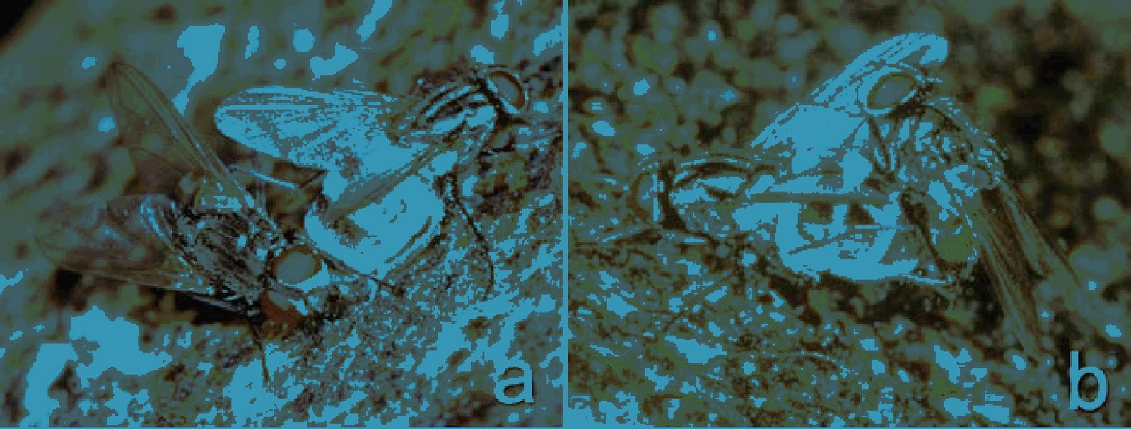
In the past, epidemics have decimated human populations. These events can directly affect human health, like the bubonic plague or indirect like the Irish potato famine. These diseases are caused by microbes, and epidemics can be just as dangerous to other species. An epidemic in fly populations caused by Entomophthora muscae is called an epizootic (1). E. muscae is a zygomycota fungus (5). This fungus infects a wide variety of fly species like, Delia antiqua, Pollenia rudis, Coenosia tigrina, the house fly (Musca domestica) along with several other hosts (1). This infection is heavily dependent on host and pathogen population densities (2). This common pathogen of flies can cause epizootics, if conditions are right, with a prevalence of 70-90%, so the majority of flies it infects die. This makes it a good biological candidate for controlling multiple fly populations. However there is some host specificity observed, E. muscae transmitted to the same type of host has a higher infection rate than those transmitted to a different type. It is unknown if this is due to genetic variation or phenotypic adaptation. The isolates from different hosts are morphologically very similar (1). Since the infection rate is dependent on fly population density it will be most effective at stopping swarms of onion flies or cabbage root flies from ruining crops. In these cases E. muscae won’t stop the maggots from damaging the plants roots, just diminish the numbers of following generations that will be present over the course of the growing season (2). Of course, the first step in this process is a fly becoming infected.

Infection by E. muscae starts with the conidia, an asexual, non-motile fungal spore, attaching to a fly. Conidia have outer mucilaginous protoplasm that they use to adhere to most substrates (2). The spores can attach and germinate at any time over the course of the day. However temperature and humidity do affect how virulent the fungus is. Higher atmospheric humidity results in more flies succumbing to the infection. Flies that survived an infection at a lower relative humidity would become lethally infected if exposed at a higher humidity (3). It doesn’t seem like the flies that fight off an infection of E. muscae become resistant to further infections. Temperature could also affect the growth rates, as E. muscae would grow slower at lower temperatures and could even be killed off at higher temperatures. Within 24 hours the spores will pierce the exoskeleton with germ tubes (4). One long germ tube arises from the secondary conidium and can branch out inside the insect hemocoel. Fungal cytoplasm will fill the germ tubes before hyphae can start growing at the ends (2, 4). Sphere like hyphae bodies start replicating in the fly within 24-48 hours. After 48 hours the hyphae start consuming the hemolymph and internal organs. Signs of an infection include changes such as swollen abdomen angled high into the air and outstretched wings. The abdomen turns white as conidiophores grow through the intersegmental membranes (6).
Figure 1. A infected female cadaver and male fly (5)
Figure 2. close up of conidiaphores (2)
It is at this point the fungus can develop two different ways each resulting in different behavior in flies. E. muscae can keep perpetuating itself through the growing season by developing conidiaophores. The infected host flies up to someplace high in the late afternoon and die a few hours later, this is know as summit disease (5). The conidiophores continue to grow and penetrate out through the membranous ventral aspect of the abdomen from between the tergites. The conidiophores are finger shaped as they start growing outwards and start resembling a bell as they mature. Only one spore is developed at the apex, which is then forcibly ejected at the onset of night (2, 4).

Figure 3. life cycle of E. muscae (5)
These conidia drift on the winds until they attach to another fly and begin the cycle again. There is another way for the conidia to find a new host. A male house fly will find an infected female cadaver that he comes across irresistible. He will try court and mate with the female and in the process become infected, even to the point of ignoring a healthy female (5, 6, 7). Another reproductive strategy of E. muscae is producing resting spores. These are primarily made in females and are dependent on the photoperiod (7). Resting spores are produced from about 10% of the infections throughout the growing season. On this path the fly is compelled to find soil to land on and perish. After the fly dies, its abdomen becomes blackened and brittle, eventually falling apart to release the resting spores into the soil (2). They will lie dormant throughout the winter and attach themselves to emerging adult flies in the early spring. Production of either spore type is greatest at 16 degrees Celsius, this is also the optimal temperature for infection. Temperature is something that the flies can exploit to increase their survivability.

Infected flies exhibit a behavior fever, where the fly will try to find hotter areas during the first few days of infection. The hosts that are able to stay in hotter areas, temperatures over 40 degrees Celsius, will have a higher survival rate (4, 5). If the flies don’t exhibit this behavior early enough it won’t be able to kill off the fungus. Such behavior is because the flies can’t efficiently regulate their body temperature. As the infection progresses the fly will seek out a cooler area and succumb to the pathogen. Some E. muscae isolates have been observed that are highly resistant to the behavioral fever. So if conditions are right it is very easy for an epizootic to occur within the fly population.

More research is currently being done to see how feasible it is to use biological controls instead of pesticides. It is true that infected flies take much longer to die than those exposed to a pesticide, but they still consume significantly less than healthy flies (5). Unlike pesticides E. muscae can be very specific and only affect a small number of species, without causing detrimental damage to the environment. The possibility of using E. muscae to keep fly populations low, near crops could be a huge boon and is worth looking into implementing.


Works Cited

1. Jensen, Annette, et al. “intraspecific variatio and host specificity of entomophthora muscae sesu stricto isolates revealed be random amplified polymorphic DNA, universal primed PCR, PCR restriction fragment length polymorphism and conidial morphology.” Invertebrate pathology 78, 251-259. January 21 2002.
2. Carruthers, RAymound, et al. “entomophthora muscae (Entomophthorales: Entomophthoracae) Mycosis in the Onion Fly, Delia antiqua (Diptera: Anthomyiidae).” Invertebrate pathology 45, 81-93. 1985.
3. Kramer, John. “THE HOUSE-FLY MYCOSIS CAUSED BY ENTOMOPHTHORA MUSCAE: INFLUENCE OF RELATIVE HUMIDITY ON INFECTIVITY AND CONIDIAL GERMINATION” new york entomological society. 88 p236-240. 1980.
4. Watson, D. et al. “behavioral fever response of musca domestica to infection by entomophthora muscae.” Invertebrate pathology 61 p10-16. 1993.
5. Roy, H. E., Steinkraus, D. C., Eilenberg, J., Hajek, a E., & Pell, J. K. (2006). Bizarre interactions and endgames: entomopathogenic fungi and their arthropod hosts. Annual review of entomology, 51, 331-57. doi:10.1146/annurev.ento.51.110104.150941
6. Zurek, L., Wes Watson, D., Krasnoff, S. B., & Schal, C. (2002). Effect of the entomopathogenic fungus, Entomophthora muscae (Zygomycetes: Entomophthoraceae), on sex pheromone and other cuticular hydrocarbons of the house fly, Musca domestica. Journal of invertebrate pathology, 80(3), 171-6. Retrieved from PubMed
7. Thomsen, L., & Eilenberg, J. (2000). Entomophthora muscae resting spore formation in vivo in the host Delia radicum. Journal of invertebrate pathology, 76(2), 127-30. doi:10.1006/jipa.2000.4961

Tuesday, December 6, 2011

Trypanosoma cruzi: The invisible killer

By Simin Golestani
For years it has been known that the protozoan parasite Trypanosoma cruzi is responsible for Chagas disease, infecting more than 18 million people worldwide, with another 120 million people at risk of acquiring this parasite in endemic regions (1). T. cruzi infection in humans occurs when reduviid bug excreta breaks into the skin, and amastigotes enter the body (2).  Once inside, the amastigotes differentiate into bloodform trypomastigotes, which specifically target muscle cells, including the heart.  The most unusual outcome of this disease is chronic Chagas disease Cardiomyopathy, CCC,  which can manifest itself up to 30 years after the initial infection with T. cruzi (1).  Patients with this disease show symptoms of myocarditis, which is a weakening of the heart muscles, ventricular enlargements, and cardiac ischemia, a lack of sufficient blood flow to the heart.  However, mysteriously, in autopsies of hearts displaying CCC there is no indication of the presence of the parasite (2).  Chagas disease cardiomyopathy is distinct from regular congestive heart failure, and the absence of this parasite in the heart leads to the hypothesis that T. cruzi’s effects on the heart are in an indirect manner.


The main reason that the mechanism of pathogeneis of Chagas disease is so difficult to understand is due to the extreme variations in the outcome of infections (1).  Acute infections manifest themselves in the form of lesions at the site of infection, and studies have shown that initially after contact, antibodies against myosin, actin and laminin have been detected in host species (2). This means that in some cases T. cruzi begins its degenerative effects on muscles immediately after infecting the host.  However, the majority of infected people can be asymptomatic for the remainder of their lives, and only one third of those infected will eventually display Chagas disease cardiomyopathy (1).  This makes early detection and treatment of CCC extremely difficult, and in the past hypothesis about the pathogenesis of Trypanosoma cruzi had been dependent solely on autopsies of infected cadaver hearts.  Recently, animal models have been used to discover many possible explanations for the secondary affects of T. cruzi on the heart.  

Figure 1. (2)  Images of an uninfected
and infected heart.  Myocyte swelling
and necrosis are observed in the
muscle tissue of the infected heart.
Years of extensive research on this topic have yet to reach a definitive conclusion as to the cause of CCC. There are currently six different explanations for T. cruzi related cardiomyopathy, and it is thought that a combination of these, not a single one, are responsible for CCC.  The presence of a specific T. cruzi secretion has not yet been demonstrated, however, it has been put forth as an explanation (1,2).  This secretion is toxic to host tissues and can continue causing damage even after the parasites are no longer present in the body.  Parasite-induced myocytolysis is a mechanism suggested for cardiomyopathy, in which the host cells lyse after the amastigotes differentiate into the trypomastigotes (2).  This will initially cause irreparable damage to the heart tissue which may not manifest itself until many years after the infection.  Parasite induced microvascualr changes could also be an explanation, depriving the heart of proper oxygen perfusion and leading to chronic inflammation.  The majority of explanations for CCC focus on autoimmunity and the ways in which this can come about, including the presence of T. cruzi antigens which can act as a site for specific T cell mediated delayed- type hypersensitivity, leading to damage of host tissues (1).  Polyclonal B cell activation is thought to occur after T. cruzi infections, leading to immunosuppresion in the host.  Bystander activation, in which T cells specific for one antigen are activated as a result of an immune response against a different antigen, is also thought to play a role in bringing about auto immune responses in Chagas disease infected patients (2).

Researchers have yet to come to a consensus about the most likely cause of autoimmunity leading to Chagas disease cardiomyopathy, however, recently the explanation receiving the most attention is that of molecular mimicry (1).  Molecular mimicry occurs when specific sequences between foreign intrusive peptides and self peptides are similar enough to result in a cross- activation of autoreactive T or B cells (4).  Occasionally, a single T cell receptor can be activated by foreign residues, which results in autoimmunity.  It is thought that the body’s immune response to a T. cruzi protein will crossreact with a self protein sharing a similar target.  This causes an immune response to be launched against the host and tissue damage occurs if this response is strong enough (1). There are two combinations hypothesized for molecular mimicry, one is the reaction of  the peptides of T cruzi B13 protein with the human cardiac myosin, and the other is the peptides of T cruzi cruzipain and skeletal myosin (1). These cross reactive T. cruzi proteins are thought to cause autoimmunity, which then results in the progressive degeneration of all muscles, especially the heart.  It has also been shown that human cardiac myosin from Chagas disease patients can specifically recognize a T. cruzi antigen (1).  It was also found that cardiac myosin antibodies were present in 100% of sera from CCC patients, but not in healthy patients.  Myosin specific autoimmunity caused in part by molecular mimicry is apparently one of the most common causes of cardiac myopathy.  
It is currently impossible to determine if any of these parasite induced effects are the sole reason for cardiomyopathy, and in reality is it mostly likely a combination of autoimmunity and anti-parasite immunity and other factors that result in eventual cardiac death (1,2). The only cure for CCC is a heart transplant, and therefore discovering the pathogenesis of T. cruzi is of extreme importance.  Until the exact mechanism of parasite induced autoimmunity is discovered, appropriate treatments will be impossible to find, and the T. cruzi killer will still be at large, claiming more than 50,000 lives each year.

Works cited:


1.  Edecio, C. Bilate, A. Hyland, K. Fonseca, S. Kalil, J. Engman, D. “Induction of cardiac autoimmunity in Chagas heart disease: A case for molecular mimicry.” Autoimmunity, 39(1): 41-54. February 2006.  


2.  Engman, D. Leon, J. “Pathogenesis of Chagas heart disease: role of autoimmunity.”
Acta Tropica 8: 123–132. October 2002.


3. Bonney, K. Engman, D. "Chagas heart disease pathogenesis: One mechanism or many?" Current Molecular Medicine, 8, 510-518. 2008.  
4. Wikepedia: molecular mimicry.  http://en.wikipedia.org/wiki/Molecular_mimicry

Monday, December 5, 2011

Pathogenic Noms for Acanthamoeba polyphaga by An Dang


             Acanthamoeba species are ubiquitous, occupying diverse habitats, including moist soils and freshwater environments.  In these natural habitats, they prey on bacteria, controlling the bacterial population, which ultimately results in the recycling of nutrients back into the ecosystem [1]. However, some bacterial species have evolved ways to evade digestion and persist within the protozoa as endosymbionts [1,2]. Acanthamoeba can withstand hostile environmental conditions such as chlorination and high temperatures. It also displays resistance to numerous disinfectants and  may exist as cysts, thus providing bacteria that may be less tolerant to environmental stresses, an enticing environment to live in [3]. Acanthamoeba are found virtually everywhere, and the ongoing interaction between protozoa and bacteria, particularly pathogenic bacterial strains are of great concern. A particular species, Acanthamoeba polyphaga, have been implicated in serving as a potential reservoir for methicillin-resistant Staphlyococcus aureus (MRSA), Campylobacter jejuni, Vibrio cholera and Escherichi coli O157. Thus, these amoeba serve as vectors for the spread of life-threatening infections [2].  Several studies have demonstrated that pathogenic bacteria species that are capable of evading digestion by amoeba can proliferate within the host and emerge as more virulent forms [4]. Therefore, it is important to study the protozoa-bacteria interactions and understand how this relationship impacts human health.
                  MRSA are able to replicate within the intracellular environment of A. polyphaga. Huws et al. 2004 have shown that in about 50% of amoebae isolated in the study, viable MRSA were detected within phago-lysosomes, and viable cocci can be detected within the cytoplasm. In a co-culture with amoeba and MRSA, extracellular numbers of bacteria were significantly larger compared to bacteria alone, demonstrating a 1000-fold difference in the number of bacteria in the presence of A. polyphaga relative to its absence. Due to the ability of protozoa to form cysts, it can also entrap MRSA and provide a means of infection via aerosilization. Therefore, this study suggests that A. polyphaga can impact the proliferation of MRSA, and be involved in its dispersal in both community and nosocomial settings [4].
                  Another study, performed by Axelsson-Olsson et al., demonstrated the protozoa’s role in supporting the life cycle of C. jejuni. Previous studies have shown that these bacteria can avoid the bactericidal effects of chlorination by residing in protozoa, and that in a co-culture, C. jejuni displayed over a 50-fold increase in resistance to chlorine compared to pure cultures. These researchers also demonstrated that these bacteria can accumulate within amoebic vacuoles and persist in the intracellular environment provided by A. polyphaga for a longer period of time when compared to co-cultures. Ultimately, the study demonstrated that C. jejuni are capable of infecting A. polyphaga, and avoid degradation by the host and proliferate intracellularly suggesting that the protozoa are capable of serving as a reservoir for C. jejuni [3]. This has important implications because infection by C. jejuni , although it is usually non-lethal, is responsible for over 2 million cases of gastroenteritis in the U.S. which can be transmitted via uncooked poultry or untreated water [5].
Vibrio cholera, particularly strains O1 and O139, causes potentially life-threatening dysentery with a 25-50% fatality rate if left untreated, is the agent responsible for the continual pandemic in South America, Africa and Asia. An individual can contract cholera by consumption of water contaminated with feces. Natural water samples obtained in Sudan, where cholera is endemic, V. cholera can be found associated with Acanthamoeba. Although it is commonly affiliated with A. castellanii, studies have demonstrated that it also interacts with A. polyphaga. These bacteria can be found within the vacuoles of the amoeba , and a co-culture demonstrated that neither species diminished the growth of the other [6]. In fact, they seemed to support each other’s growth, in that 89% of V. cholera were found associated with Acanthamoeba. By residing in Acanthamoeba, V. cholera can escape the selective pressures of chlorination and even the actions of antibiotics and contribute to the severity of human illness. Thus, it is important to understand the interactions between these two organisms to develop an efficient way to combat the spread of disease [6,7].
                  Escherichia coli O157:H7 are responsible for haemolytic uraemic syndrome outbreaks in North America, and this strain is highly transmissible, and only requires ingestion of about 100 bacterial cells. In 1997, mud contaminated with E. coli O157 may have been the source of the outbreak, suggesting that an environmental source may be responsible for its continual recycling in the environment causing re-current infections in cattle. Studies suggest that a possible source may be ubiquitous organisms, such as amoeba like A. polyphaga. These protozoa could prey on E. coli and use it as a food source, but there are cases where E. coli can escape digestion and proliferate within vacuoles. However, the exact mechanism of how certain E. coli evade digestion remains unknown. In any case, studies suggest that because E. coli can live within Acanthamoeba, the amoeba may serve as a vector for possible E. coli infections [8].
                  Although Acanthamoeba species are more commonly associated with amoebic infections such as keratitis and potentially fatal granulomatous encephalitis, its role as a potential reservoir for certain pathogenic bacteria have been overlooked.  Their ability to inhabit diverse areas and resist environmental stresses makes them very attractive hosts for bacteria which are less tolerant.  It remains to be discovered the mechanisms of how certain bacteria are able to evade digestion and proliferate within the intracellular environment by the amoeba, and how it provides a selective environment for more pathogenic strains. However, its ability to play host to a plethora of bacterial organisms has important implications in the spread and maintenance of disease both in nosocomial and community settings.


Works Cited

1.            Barker J, Brown MRW (1994) Trojan Horses of the microbial world: protozoa and the survival of bacterial pathogens in the environment. Microbiology 140: 1253–1259. Available:http://mic.sgmjournals.org/cgi/doi/10.1099/00221287-140-6-1253. Accessed 10 November 2011.
2.            Huws SA, Morley RJ, Jones MV, Brown MRW, Smith AW (2008) Interactions of some common pathogenic bacteria with Acanthamoeba polyphaga. FEMS microbiology letters 282: 258–265. Available:http://www.ncbi.nlm.nih.gov/pubmed/18399997. Accessed 13 August 2011.
3.            Axelsson-Olsson D, Waldenström J, Broman T, Olsen B, Holmberg M (2005) Protozoan Acanthamoeba polyphaga as a potential reservoir for Campylobacter jejuni. Applied and environmental microbiology 71: 987–992. Available:http://www.pubmedcentral.nih.gov/articlerender.fcgi?artid=546671&tool=pmcentrez&rendertype=abstract. Accessed 10 November 2011.
4.            Huws SA, Smith AW, Enright MC, Wood PJ, Brown MRW (2006) Amoebae promote persistence of epidemic strains of MRSA. Environmental microbiology 8: 1130–1133. Available:http://www.ncbi.nlm.nih.gov/pubmed/16689734. Accessed 10 November 2011.
5.            Campylobacter jejuni (2011). Available:www.cdc.gov. Accessed 11 October 2011.
6.            Sandström G, Saeed A, Abd H (2010) Acanthamoeba polyphaga is a possible host for Vibrio cholerae in aquatic environments. Experimental parasitology 126: 65–68. Available:http://www.ncbi.nlm.nih.gov/pubmed/19815016. Accessed 12 November 2011.
7.            Shanan S, Abd H, Hedenström I, Saeed A, Sandström G (2011) Detection of Vibrio cholerae and Acanthamoeba species from same natural water samples collected from different cholera endemic areas in Sudan. BMC research notes 4: 109. Available:http://www.pubmedcentral.nih.gov/articlerender.fcgi?artid=3080310&tool=pmcentrez&rendertype=abstract. Accessed 12 November 2011.
8.            Barker J, Humphrey TJ, Brown MW (1999) Survival of Escherichia coli O157 in a soil protozoan: implications for disease. FEMS microbiology letters 173: 291–295. Available:http://www.ncbi.nlm.nih.gov/pubmed/10227158. Accessed 12 November 2011.

Paracoccidioidomycosis: Men And Women Are Not Created Equal.

Figure 1. Skin lesions on a man infected with P. brasiliensis.Image from NetHealthBook.com    
Paracoccidioidomycosis (PCM) is caused by a human fungal pathogen primarily infecting the lungs through inhalation of pathogenic conidia and can then disseminate throughout the rest of the body resulting in lesions in the skin, lymph nodes, mucous membranes, and adrenals (see Figure 1). It is the most common mycosal infection in Latin America, caused by the fungi Paracoccidioides brasiliensis. After inhalation, single nucleus conidia are transformed into multinucleated yeast at human temperature [4]. The natural habitat of P. brasiliensis is still unknown, but a case study by Calle et al. characterized environmental conditions in PCM endemic areas and found that a large number of endemic areas are located in coffee-growing areas in Colombia [3]. One of the most interesting clinical observations made about PCM is that incidence of symptomatic infection is not equal between males and females. There have been reports varying from a 13:1 up to 70.6:1 ratio of men-to-women incidence of infection [10]. What is the cause of this very extreme difference in infection rate between genders? Is it an environmental effect or is a physiological difference responsible for this huge difference in rate of infection?

Because P. brasiliensis’ natural habitat may be in close proximity to coffee plantations in Colombia and possibly other agricultural areas in Latin America, an initial hypothesis of the phenomenon that men contract PCM much more frequently than females may be that men interact more with the fungus through agriculture and therefore contract infection more often. However, skin tests in endemic areas have shown equal reaction to the fungal polysaccharide antigen, paracoccidioidin, indicating an equal presence of P. brasiliensis in males and females, suggesting that a biological difference between genders plays a role in the pathogenicity of Paracoccidioidomycosis [1, 8].

It is known that in order for initial infection to occur, P. brasiliensis must transition from its mycelial form to yeast form [8,9]. A study by Restrepo et al. found that the female sex hormone estrogen (specifically 17β-estradiol), and no other steroid hormones, suppresses this transition in vitro. P. brasiliensis isolates were also found to express a protein which binds estrogen, and is believed to be the molecular site of action for transition disruption [8]. This explains why paracoccidioidin skin tests on females return positive in equal frequency to men even when females are asymptomatic; the fungal cell is arrested in its mycelial form, preventing the spread of infection. Aristizabal et al. demonstrated these effects in vivo by castrating male and female mice and reconstituting some of the castrated males with β17-estradiol and some of the castrated females with testosterone. When infected with P. brasiliensis, normal female mice are able to control infection while normal males experience progressive disease. Castrated males, castrated females, and castrated females reconstituted with testosterone were unable to control infection, but castrated males reconstituted with β17-estradiol were able to restrict fungal multiplication [2]. This study shows the important role of β17-estradiol in host immunity that testosterone does not act equivalently.

This brings the question of how estrogen disrupts the morphological switch of P. brasiliensis and contributes to the host immune response during infection. It has been found that estrogen specifically binds to mycelial cytosolic extracts of P. brasiliensis, and the fungal binder is likely a protein that acts as an estradiol binding protein. Study of temporal gene expression during the morphological change from mycelium to yeast form demonstrated that treating mycelia with estrogen caused differential gene expression compared to controls and cells were arrested in the mycelial form [10]. Also, higher levels of the cytokines IL-2, IFN-γ, and TNF-α have been correlated with resistance to infection, and higher levels of IL-10 play a major role in the antigen-specific immunosuppression of PCM. Likewise, estrogens have been found to stimulate IL-12, IFN-γ, and TNF-α and down regulate IL-10, while testosterone is known to actually increase synthesis of IL-10 [7]. Estrogen’s ability to stimulate cytokines necessary to fight infection while down regulating cytokines with immunosuppressive activity may therefore contribute to females’ ability to resist PCM. It may also suggest that testosterone actually contributes to the male hosts’ susceptibility to infection with increased production of IL-10.

PCM isn’t the only microbial infection found to be more prevalent in males than females. Gastric cancer risk is two times greater in males than females, and one of the causes is from Helicobacter pylori colonized in the gastric epithelia [6]. H. pylori infected mice treated with 17β-estradiol have been found to develop less severe symptoms than control male mice, suggesting a similar effect in host immunity during H. pylori infection [5]. In contrast to P. brasiliensis and H. pylori, it’s been found that Candida albicans has an estrogen-binding protein with a high affinity for estradiol, which actually stimulates the dimorphic transition from yeast the hyphal form [10].

When it comes to PCM women fair better than men, and they have estrogen to thank for it. 17β-estradiol blocks the critical morphological transition of P. brasiliensis from mycelial to yeast form through stereospecific binding to fungal proteins and functions in the regulation of immune system cytokines. Though estrogen has been found to be beneficial in host immunity, it is also harmful during certain pathogenic infections. Sex hormones play an important role in host immunity and further study of their mechanisms of action may offer helpful insights in the battle against human pathogens.

by RH

References:
1. Andc, M. O. K., Netto, F., Sciences, M., Abstract, B., Basin, U. A., America, L., Accepted, S., et al. (1978). PARACOCCIDIOIDIN AND HISTOPLASMIN IN COARI (STATE OF AMAZONAS), SENSITIVITY. Tropical Medicine, 27(4), 808-814.
2. Aristizábal, B. H., Clemons, K. V., Cock, a M., Restrepo, a, & Stevens, D. a. (2002). Experimental paracoccidioides brasiliensis infection in mice: influence of the hormonal status of the host on tissue responses. Medical mycology: official publication of the International Society for Human and Animal Mycology, 40(2), 169-78.
3. Calle, D., Rosero, D. S., Orozco, L. C., Camargo, D., Castañeda, E., & Restrepo, a. (2001). Paracoccidioidomycosis in Colombia: an ecological study. Epidemiology and infection, 126(2), 309-15.
4. Goldani, LZ.(2011). Gastrointestinal Paracoccidioidomycosis: An Overview. J Clin Gasteroenterol 45(2), 85-91.
5. Ohtani, M., Ge, Z., García, A., Rogers, A. B., Muthupalani, S., Taylor, N. S., Xu, S., et al. (2011). 17 β-estradiol suppresses Helicobacter pylori-induced gastric pathology in male hypergastrinemic INS-GAS mice. Carcinogenesis, 32 (8), 1244-50.
6. Parkin, D.M., et al. (2005). Global cancer statistics, 2002. CA Cancer J. Clin., 55, 74-108.
7. Pinzan, C. F., Ruas, L. P., Casabona-Fortunato, A. S., Carvalho, F. C., & Roque-Barreira, M.-C. (2010). Immunological basis for the gender differences in murine Paracoccidioides brasiliensis infection.
8. Restrepo, a, Salazar, M. E., Cano, L. E., Stover, E. P., Feldman, D., & Stevens, D. a. (1984). Estrogens inhibit mycelium-to-yeast transformation in the fungus Paracoccidioides brasiliensis: implications for resistance of females to paracoccidioidomycosis. Infection and immunity, 46(2), 346-53.
9. Rippon, J. W. (1980). Dimorphism in pathogenic fungi. Critical reviews in microbiology, 8(1), 49-97. 
10. Shankar, J., Restrepo, A., Clemons, K. V., & Stevens, D. a. (2011). Hormones and the resistance of women to paracoccidioidomycosis. Clinical microbiology reviews, 24(2), 296-313.